IN THE PRESS

Chirurgie du cancer : une sonde pour améliorer le pronostic en cancer du cerveau et du sein
Une sonde chirurgicale développée à Polytechnique Montréal est en voie de prendre sa place dans les blocs opératoires de la planète.

Une sonde innovante pour mieux opérer les cancers
En exploitant les propriétés de la lumière, une équipe a réussi à repérer les cellules cancéreuses, en pleine opération du cerveau. Une technologie très prometteuse.

Percée scientifique sur les tumeurs cérébrales
Une nouvelle technique permettant de détecter les tissus cancéreux, dans certains types de tumeurs au cerveau, fait maintenant son apparition dans la pratique médicale. Des chercheurs d’instituts québécois ont développé une sonde permettant ce genre de pratique.

Visualizing secrets of glioma
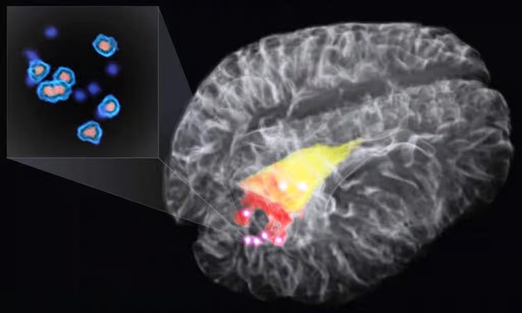
A new approach developed by Frederic Leblond, Kevin Petrecca and colleagues offers improved definition of cancer and normal tissues during surgery. The Raman spectroscopy modality distinguishes tissues based on their different molecular characteristics, which result in variable inelastic scattering of incident laser light and thus distinct spectral profiles. “This handheld Raman spectroscopy probe technique, coupled with our machine-learning tissue-classification algorithms, can detect brain cancer and, most importantly, invasive cancer cells on a background of normal brain, at cellular resolution,” says Leblond.

L’effet Raman, un nouvel outil contre le cancer du cerveau
Le cancer du cerveau est particulièrement difficile à traiter, en particulier le glioblastome qui a un taux de survie faible. Une nouvelle technique basée sur l’effet Raman permet désormais aux neurochirugiens de repérer plus efficacement lors d’une opération les cellules cancéreuses du cerveau. De quoi améliorer le pronostic de survie des patients.

New Raman Spectroscopy Probe Could Improve Outcomes In Brain Cancer Surgery
Scientists have developed a new intraoperative probing technique that could increase survival odds for patients with brain cancer. The McGill University researchers claim that a Raman spectroscopy probe can be used during surgery to accurately identify almost all invasive brain cancer cells that other methods could potentially miss.

New laser probe identifies brain cancer cells in real time
Promises to improves tumor surgeries and extend survival times for brain cancer patients
A new intraoperative handheld probe for cancer-cell-detection enables surgeons, for the first time, to detect more than 92% of invasive brain cancer cells in real time during surgery, according to its developers at the Montreal Neurological Institute and Hospital – The Neuro, McGill University MUHC, and Polytechnique Montréal.

Light-Based Technique Helps Surgeons Excise Brain Cancer
Neurosurgeons need all the help they can get to remove brain cancer tumors. If they leave cancer cells behind, the tumors can regrow. Finding cancer cells can be particularly difficult with infiltrative cancers such as glioma, which invades surrounding brain tissue.

New Techniques Outline Tumors' Location in the Brain
Brain tumors are notoriously tricky for surgeons, who may leave too much cancerous tissue behind or cut into vital, healthy brain tissue.
However, two new studies describe devices that help surgeons and nonsurgical physicians better understand the outline and location of cancerous tissue in the brain, potentially improving outcomes for patients.

Handheld Probe Detects Cancer Cells in Brain
Medical and engineering researchers at McGill University and Polytechnique Montréal developed a technique using a handheld probe that enables surgeons to find elusive cancer cells in the brain. The team led by McGill’s Kevin Petrecca and Polytechnique Montréal’s Frederic Leblond reported their findings yesterday in the journal Science Translational Medicine (paid subscription required).

New Handheld Probe Detects Cancer Invaders in the Brain
Some cancers appear indistinguishable from healthy tissue, raising the risk of recurrence and metastasis. Now, researchers in Canada have developed a handheld fiber optic probe that they say can detect invasive brain cancer in patients accurately. The device, described in Science Translational Medicine this week, could one day quickly guide neurosurgeons during procedures to remove cancerous tissue from the brain.

Revolutionary new probe zooms in on cancer cells
Brain cancer patients may live longer thanks to a new cancer-detection method developed by researchers at the Montreal Neurological Institute and Hospital - The Neuro, at McGill University and the MUHC, and Polytechnique Montréal. The collaborative team has created a powerful new intraoperative probe for detecting cancer cells. The hand-held Raman spectroscopy probe enables surgeons, for the first time, to accurately detect virtually all invasive brain cancer cells in real time during surgery. The probe is superior to existing technology and could set a new standard for successful brain cancer surgery.

Revolutionary new probe zooms in on cancer cells
Improves tumour surgeries and extends survival times for brain cancer patients
Brain cancer patients may live longer thanks to a new cancer-detection method developed by researchers at the Montreal Neurological Institute and Hospital – The Neuro, at McGill University and the MUHC, and Polytechnique Montréal. The collaborative team has created a powerful new intraoperative probe for detecting cancer cells. The hand-held Raman spectroscopy probe enables surgeons, for the first time, to accurately detect virtually all invasive brain cancer cells in real time during surgery. The probe is superior to existing technology and could set a new standard for successful brain cancer surgery.

Raman Technique Helps Surgeons Excise Brain Cancer
Neurosurgeons need all the help they can get to remove brain cancer tumors. If they leave cancer cells behind, the tumors can regrow. Finding cancer cells can be particularly difficult with infiltrative cancers such as glioma, which invades surrounding brain tissue.

De l’infiniment grand à l’infiniment petit
C’est l’histoire d’une belle aventure scientifique, celle d’un jeune chercheur québécois passionné d’astrophysique qui a conçu une caméra ultrasensible permettant de détecter les photons émis par des exoplanètes situées à des années-lumière de nous. Mais le plus étonnant est que cette technologie qu’il a imaginée pour sonder l’infiniment grand sert aussi à voir l’infiniment petit, comme des cellules cancéreuses enfouies dans le cerveau.

Nüvü Camēras Supports Medicine By Offering The Most Sensitive Brain Cancer Treatment
With the contribution made by Yoann Gosselin, recent graduate in Biomedical Engineering at l’École Polytechnique de Montréal, brain cancer treatment is leaping forward. Taking advantage of Nüvü Camēras’ HNü unique performances, he significantly heightened the sensitivity of a technique used in the removal of cancerous cells in the brain, renowned as being the most effective to this day. Combined with radiotherapy and/or chemotherapy, this novel treatment will provide a better prognosis for brain cancer patients whose current life expectancy remains very limited.

Applications Expand for Photon Counting
The prospects of photon-counting imaging are as diverse as they are promising. From space-debris monitoring, to telecommunication satellites and shuttle missions, to the advent of powerful surgical tools, which could help cancer patients through the precise removal of malignant tissues, its applications are numerous and expanding.
Photon counting reveals its strengths in extremely low-light conditions, where other imaging techniques fail to provide valuable data. This is true for all things infinitely distant or infinitely small.
